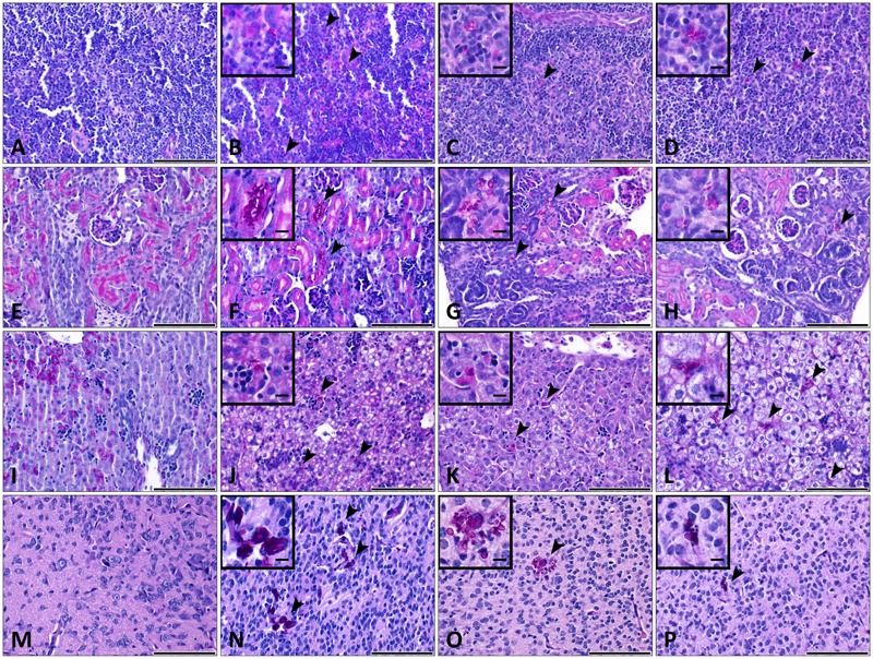
FIGURE 8.

Histopathology during systemic WT CLIB, och1Δ/Δ, and och1Δ/Δ+OCH1 C. parapsilosis infection in neonate mice. Sections were stained with periodic acid-Schiff (PAS) stain. Fungal cells (indicated by arrowheads) are evident in the organs of mice infected with the C. parapsilosis CLIB 214 (WT CLIB) (B spleen, F kidney, J liver, N brain), the och1Δ/Δ (C spleen, G kidney, K liver, O brain) and the och1Δ/Δ+OCH1 (D spleen, H kidney, L liver, P brain) strain. (A spleen, E kidney, I liver, M brain) Control organs from PBS-injected mice. All observations of the infected organs were performed at day 2 of 2 × 10⁷/20 μl dose of infection. Scale bar represents 100 μm.
