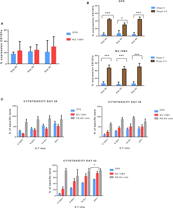
Figure 4.
(A) Expression of CD107a, a marker of degranulation, in the in vitro-generated natural killer (NK) cells (CD56+) at different time points of the differentiation protocol in response to the stimulation with K562 target cells. (B) Expression of CD107a in both conditions (with OP9 and M2-10B4 feeder cell layers) at stage 3 and stages 4–5 at different times of the differentiation protocol. (C) Cytotoxicity activity of NK cells against K562 target cells at days 28, 35, and 42 of the differentiation protocol. Overnight cultured NK cells obtained from adult peripheral blood stimulated with the same cytokines as our in vitro-generated NK cells were used as a control. The bars represent the mean and error bars represent SEM. p-Value: *p < 0.05; ***p < 0.001. The absence of any asterisk indication means there are no significant differences (Stage <3: CD56−, CD94−, CD117low; Stage 3: CD56+, CD94−, CD16−, CD117high; Stage 4: CD56+, CD94+, CD16−, CD117low, and Stage 5: CD56+, CD94+, CD16+, CD117low).

