. 2017 Jun 26;26(3):158–167. doi: 10.5607/en.2017.26.3.158

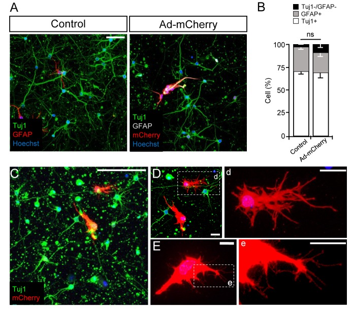
Fig. 2. Ad-CMV-mCherry infection induces cellular hypertrophy of astrocytes in 3D culture system. (A, B) Neuron-astrocyte 3D co-cultures were infected with Ad-CMV-mCherry at DIV 3 and fixed at DIV 7, followed by immunostaining with anti-TuJ1 and anti-GFAP antibodies. Nuclei are stained with Hoechst. Control indicates intact cultures that were not infected with any virus. Representative images (A) and quantification (B) of neurons and astrocytes from six independent experiments are shown. (C~E) Representative images of a neuron-astrocyte 3D co-culture infected with Ad-CMV-mCherry at DIV 3 and fixed at DIV 7. Note the cellular hypertrophy of astrocytes induced by viral infection. D and E, enlarged images of astrocytes expressing mCherry in C. Astrocyte image is rotated in E. Boxed regions in D and E are further enlarged in right (d and e) to clearly show the processes from reactive astrocytes. Scale bars: 50 µm in A, 100 µm in C, 20 µm in D, and 10 µm in E.

Fig. 2