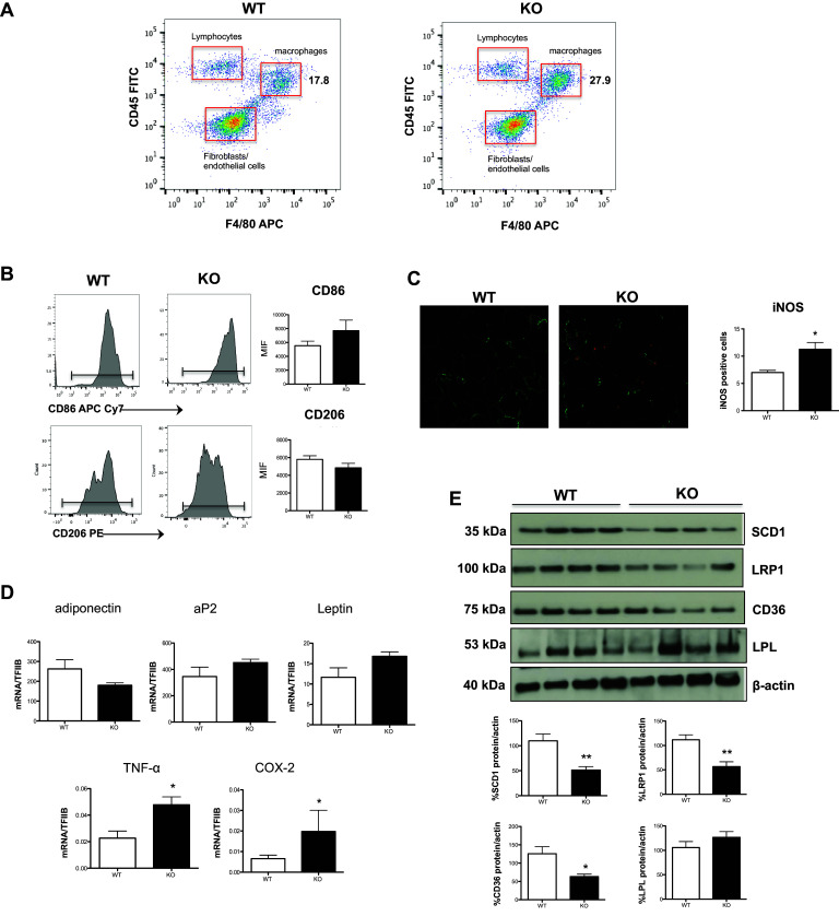
Fig. 4.

Analysis of inflammatory and adipogenic state of white adipose tissue. SVF was isolated from WAT of both WT and Elovl2−/− mice. a Gating strategy to analyze the amount of macrophages (F4/80) present in the cell fraction excluding lymphocytes (F4/80-CD45+) and fibroblast and endothelial cells (F4/80-CD45−). b CD206 and CD86 amount by flow cytometry upon staining cells at cell surface. Data are reported as mean fluorescence intensity (MFI), and are representative of three independent experiments. c Immunofluorescence of iNOS was performed by confocal laser-scanning microscopy, and data are shown as pictures taken with a Plan Fluor 40× Oil objective and as analysis (mean ± SD) of five independent experiments. d mRNA analysis of adiponectin, aP2, leptin and pro-inflammatory markers TNF-α and COX-2 in WAT. Data are shown as mean (±SD) of four independent experiments, each in duplicate. e Western blot analysis of SCD1, LRP1, CD36 and LPL, with the expected molecular mass of each protein shown on the left-hand side. Data are expressed in comparison with β-actin, and are the mean (±sem) of four different mice. *p < 0.05 versus WT; **denotes p < 0.01 versus WT
