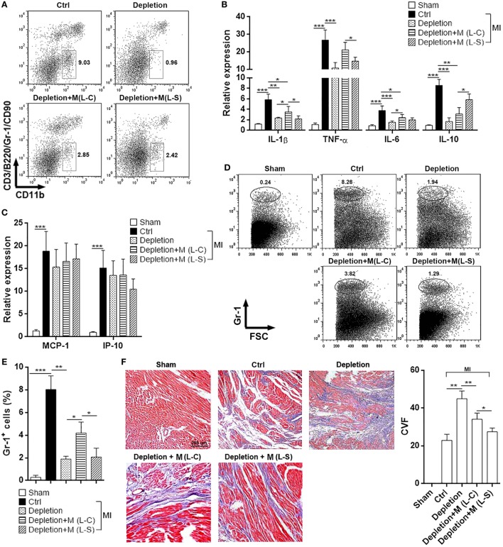
Figure 5.
Ectopic Sestrin2 expression in M1 macrophages attenuates post-MI inflammation and favors tissue repair in hearts. (A) Macrophage abundance in infarcted myocardium at day 3 after myocardial infarction (MI) (day 4 after Clophosome® treatment). Ctrl: un-pretreated mice. Depletion: Clophosome® injection. Depletion + M (L-C): Clophosome® injection followed by transfer of M1 macrophages transduced with control lentivirus. Depletion + M (L-S): Clophosome® injection followed by transfer of M1 macrophages transduced with SESN2 lentiviral activation particles. Numbers in the plots are proportions of gated cells populations. This is a representative of two independent experiments. (B,C) mRNA abundance of indicated cytokines and chemokines in infarcted myocardium. (D,E) Percentage of Gr-1+ neutrophils in the whole cardiac cells at day 3 after MI. Representative flow cytometry dot plots are shown in (D), and statistics is shown in (E). (F) Masson staining at day 7 after MI. Left panel: representative images. Right panel: statistics of collagen volume fraction (CVF). N = 4–5 per group. *p < 0.05; **p < 0.01; ***p < 0.001.

