Figure 1.
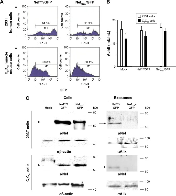
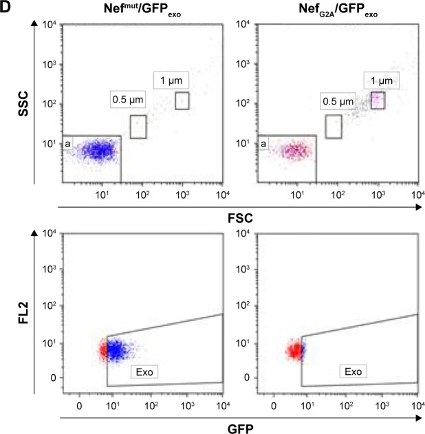
Detection of engineered exosomes in supernatants of transfected murine muscle cells.
Notes: (A) FACS analysis of both human 293T and murine C2C12 muscle cells 2 days after transfection with either Nef mut/GFP- or NefG2A/GFP-expressing vectors. M1 marks the range of positivity as established by the analysis of mock-transfected cells. Percentages of positive cells are reported. (B) Quantification in terms of AchE activity of exosome preparations recovered by differential centrifugations of supernatants from the same number (ie, 5×10⁶) of both 293T and C2C12 transfected cells. (C) Western blot analysis of exosomes from both 293T and C2C12 transfected cells. Nef-based products were detected in both cell lysates and exosomes, while β-actin and Alix served as markers for cell lysates and exosomes, respectively. Arrows mark the relevant protein products. Molecular markers are given in kDa. (D) FACS analysis of exosomes from C2C12 transfected cells. Ten mU of exosomes from C2C12 cells transfected with either Nefmut/GFP- or NefG2A/GFP-expressing vectors were analyzed in terms of both FSC and SSC (top panels), as well as GFP fluorescence (bottom panels). Quadrants indicate the dimension of the detected particulate (top panels, a: 0.1 μm) and the range of positivity as calculated by the analysis of exosomes from mock-transfected cells (bottom panels). Results are representative of two independent experiments.
Abbreviations: AchE, acetylcholineesterase; exo, exosomes; FACS, fluorescence-activated cell sorting; FL2, fluorescence channel 2; FSC, forward scatter; GFP, green fluorescent protein; SSC, side scatter.


