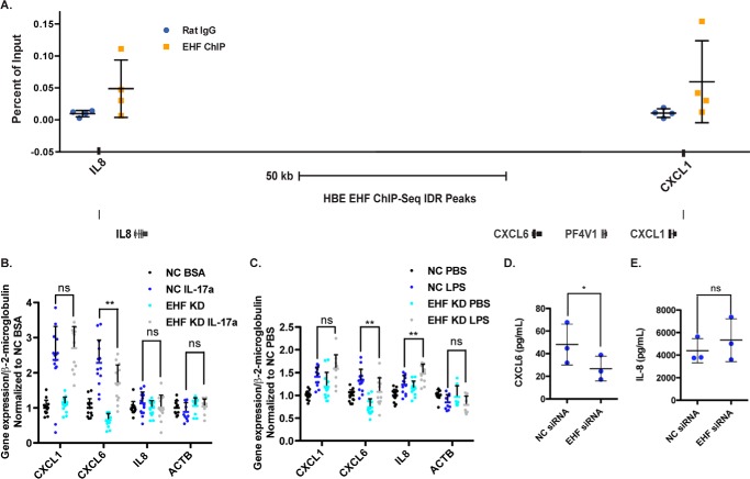
Figure 5.
EHF depletion in Calu-3 cells alters secretion of a neutrophil chemokine. A, EHF ChIP qPCR confirmed its binding to sites near IL8, CXCL6, and CXCL1 in HBE cells. Shown is the average of all experiments with standard deviation (n = 4) (top panel). Also shown is a graphic of the Chr4q13.3 region with EHF ChIP-seq peaks (bottom panel). B and C, EHF was depleted (siRNA) in Calu-3 cells, followed by their exposure to carrier, IL-17a (B), or LPS (C). Gene expression was measured by RT-qPCR. β-actin (ACTB) was included as a negative control. **, p < 0.01 by two-way analysis of variance plus multiple comparisons test; ns, not significant (average with standard deviation). D and E, secretion of CXCL6 (D) and IL-8 (E) into medium conditioned by NC- and EHF-depleted Calu-3 quantified by colorimetric sandwich ELISA (n = 3). *, p < 0.05; paired two-tailed Student's t test. Bars show the average of all experiments, with error bars representing standard deviation.

