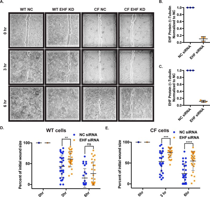
Figure 6.
EHF depletion slows wound closure in WT and CF HBE cells. A, images of NC and EHF siRNA-treated WT and CF undifferentiated HBE cells 0, 3, and 6 h after initial wounding. Outlines of wounds are traced in white. B and C, siRNA depletion of EHF was confirmed by Western blot and quantified using gel densitometry in WT (B) and CF cells (C) (average with standard deviation). D and E, percent of initial wound size was measured at all time points. Shown is the average with standard deviation for WT (D) and CF HBE cells (E) (three donor cultures). **, p < 0.01; ***, p < 0.001; ****, p < 0.0001; NS, not significant; unpaired two-tailed Student's t test. Bars show the average of all experiments, with error bars representing standard deviation.

