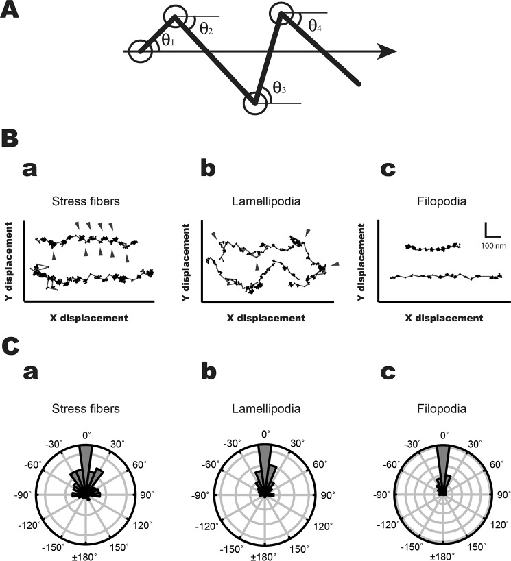
Figure 5.
Stepping orientation of human myosin VIIa movements in MEF-3T3 cells. A, diagram of measurement of stepping orientation. The arrow shows the direction of the movement. The HM7AΔTail/LZ stepping angles (θ1, θ2, θ3, θ4, … θn) were defined by the stepping angle of fluorophore movement to the axis of the movement. B, the typical stepping traces of the movement in stress fibers (a), lamellipodia (b), and filopodia (c). The arrowheads indicate the lateral positions in stress fibers and lamellipodia. The HM7AΔTail/LZ frequently shows off-axis on stress fibers, whereas the direction of HM7AΔTail/LZ on filopodia is mostly straight. The movement of HM7AΔTail/LZ on lamellipodial actin was diverse. The scales of x and y axes are shown in c. C, polar plots for orientation of HM7AΔTail/LZ movement on stress fibers (a), lamellipodia (b), and filopodia (c). The stepping orientation of HM7AΔTail/LZ was measured and plotted as described under “Experimental Procedures.” The 0° means that the stepping orientation is parallel to the moving direction.

