Figure 4.
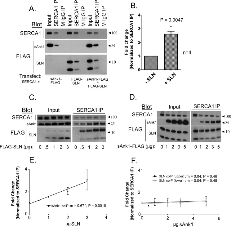
SLN promotes interaction between SERCA1 and sAnk1. A, COS7 cell extracts transfected as indicated below each panel were subjected to IP with antibodies specific to SERCA1. Transfections were performed using a [C]final of 1 μg/ml. B, quantitative densitometry analysis was performed to assess coIP between SERCA1 and sAnk1 in the presence or absence of FLAG-SLN. There was a 2.6-fold increase in coIP of sAnk1 when it was coexpressed with FLAG-SLN. C, increasing the amount of cDNA encoding FLAG-SLN, as indicated below each panel, increased interaction between SERCA1 and sAnk1. D, increasing the amount of cDNA encoding sAnk1-FLAG had no effect on coIP of SERCA1 and FLAG-SLN. E and F, graphical representation of densitometric analysis of experiments shown in C and D, respectively. Linear regression shows a significant increase in coIP of sAnk1 with SERCA1 with increasing SLN expression (E: m = 0.672 ± 0.15; p = 0.0018; n = 2), whereas increasing sAnk1 expression had no significant effect on coIP of SLN with SERCA1 (F: m = 0.0404 ± 0.53; p = 0.46 (upper band) and m = 0.040 ± 0.05080; p = 0.45 (lower band; n = 2). Data represent slope ± S.E.

