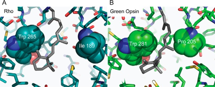
Figure 5.

Comparison of the Rho and green cone pigment-binding pockets. A, in the Rho structure, 11-cis-retinal is flanked on either side by Trp-265 and Ile-189. B, predicted green cone pigment binding site shows Pro-205, which is less flexible than the corresponding Ile-189 residue in Rho, near the chromophore.
