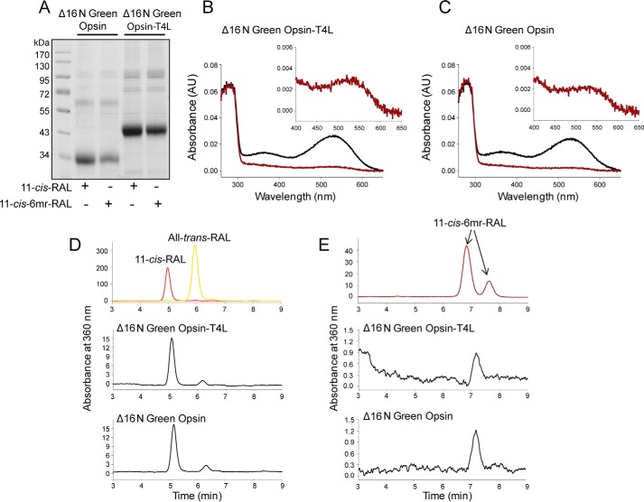
Figure 8.
Binding of 11-cis-retinal and its locked 11-cis-6mr-retinal analogue to green cone opsin lacking its N-terminal 16 amino acids. A, SDS-polyacrylamide gel of regenerated and purified pigments, Δ16N green cone opsin-T4L and Δ16N green cone opsin. Pigment regeneration was carried out in membranes isolated from insect cells, which then were purified by 1D4 immunoaffinity chromatography. Two μg of protein were loaded on each lane of the SDS-polyacrylamide gel and stained with Coomassie Blue. Proteins were deglycosylated with PNGase F before loading onto the gel. B, UV-visible absorption spectra of reconstituted Δ16N green cone opsin-T4L. C, UV-visible absorption spectra of reconstituted Δ16N green cone opsin. Spectra of pigments regenerated with 11-cis-retinal are colored black, and those regenerated with 11-cis-6mr-retinal are colored red. D and E, HPLC analyses of retinoid oximes in Δ16N green cone opsin regenerated with 11-cis-retinal and 11-cis-6mr-retinal, respectively. Retinoids were identified based on their order of elution from an HPLC column compared with that of authentic standards. D, top panel, HPLC elution profile of 11-cis-retinal and all-trans-retinal. E, top panel, HPLC elution profile of 11-cis-6mr-retinal. D and E, middle panel, elution profile of retinoid extracted from Δ16N green cone opsin-T4L regenerated with 11-cis-retinal and 11-cis-6mr-retinal, respectively. D and E, bottom panel, retinoid elution profile extracted from Δ16N green cone opsin regenerated with 11-cis-retinal and 11-cis-6mr-retinal, respectively.

