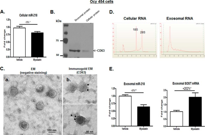
Figure 2.
Myostatin inhibited miR-218 expression in Ocy454 parent cells and their exosomes. As shown in Fig. 1A, Ocy454 cells were differentiated for 12 days and then treated with 100 ng/ml myostatin or vehicle for 48 h followed by extraction of total RNA from Ocy454 parent cells or the isolation of exosomes released form Cyc454 cells. A, levels of cellular miR-218 in Ocy454 parent cells were determined by real-time PCR. B, the Western blot showed that CD63 proteins are enriched in isolated exosome compared with that in cellular protein lysate. C, electron microscopy analysis of exosomes; a, exosomes were stained with uranyl acetate (EM (negative staining)); b, exosomes were labeled with 10-nm immunogold using antibody against exosomal membrane marker CD63 and stained with uranyl acetate (Immunogold EM (CD63)). Exosome morphology was then visualized using electron microscope Hitachi H7000. D, isolated cellular or exosomal RNA was analyzed using a Bioanalyzer. The results showed the absence of the ribosomal RNA peaks (18S and 28S rRNA) in exosomal RNA compared with that of cellular RNA. E, levels of miR-218 and SOST mRNA in exosomes produced by Ocy454 cells were determined by real-time PCR. Data shown are mean values ± S.E. for three separate determinations. *, p < 0.05 and **, p < 0.01.

