Figure 3.

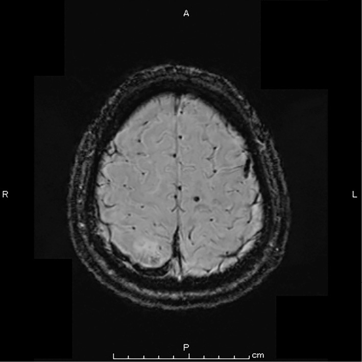
Susceptibility weighted imaging (SWI) revealed a lesion with a low signal intensity in the right cerebral cortical veins, and enlargement indicating cerebral venous thrombosis.

Susceptibility weighted imaging (SWI) revealed a lesion with a low signal intensity in the right cerebral cortical veins, and enlargement indicating cerebral venous thrombosis.