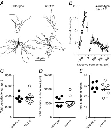
Figure 6. Dendritic morphology is not different between WT and fmr1−/y L2/3 neurons.

A, representative Neurolucida reconstructions of L2/3 pyramidal neurons from wild‐type and fmr1−/y mice showing dendritic branching patterns (wild‐type: 7 cells/7 mice; fmr1−/y: 8 cells/8 mice). B, Sholl analysis plot showing no significant difference in dendritic branching pattern between wild‐type and fmr1−/y L2/3 neurons. C–E, summary graphs showing no significant difference in dendritic length, surface area, or number of dendritic branch points between wild‐type and fmr1−/y L2/3 neurons.
