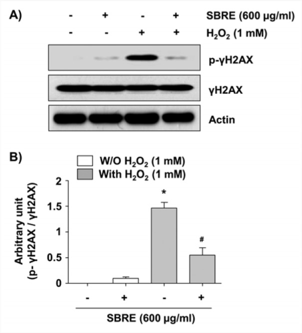
Figure 4. Attenuation of H2O2-induced phosphorylation of γH2AX by SBRE in HaCaT keratinocyte cells. Cells were pretreated with 600 μg/ml SBRE for 1 h and then stimulated with or without 1 mM μM H2O2 for 24 h. (A) The cells were lysed and then equal amounts of cell lysates were separated on SDS-polyacrylamide gels and transferred to PVDF membranes. The membranes were probed with specific antibodies against γH2AX and p-γH2AX, and the proteins were visualized using an ECL detection system. Actin was used as an internal control. (B) The relative expression of p-γH2AX represents the average densitometric analyses as compared with γH2AX. Each point represents the mean ± SD of three independent experiments (*P < 0.05 vs. untreated control; #P < 0.05 vs. H2O2-treated cells).

