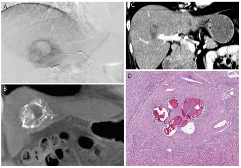
Figure 5.
A leiomyosarcoma metastasis to the liver was embolized with 100–300 μm, doxorubicin-eluting microspheres to reduce tumor size and vascularity prior to surgical resection. Selective catheterization (A) of the tumor was performed, with proper positioning confirmed by cone-beam CT (B). Post-procedure CT (C) demonstrates no residual vascularity within the lesion. Following liver resection, microspheres were identified within the blood vessels in the tumor bed without any evidence of viable metastatic tumor (D).

