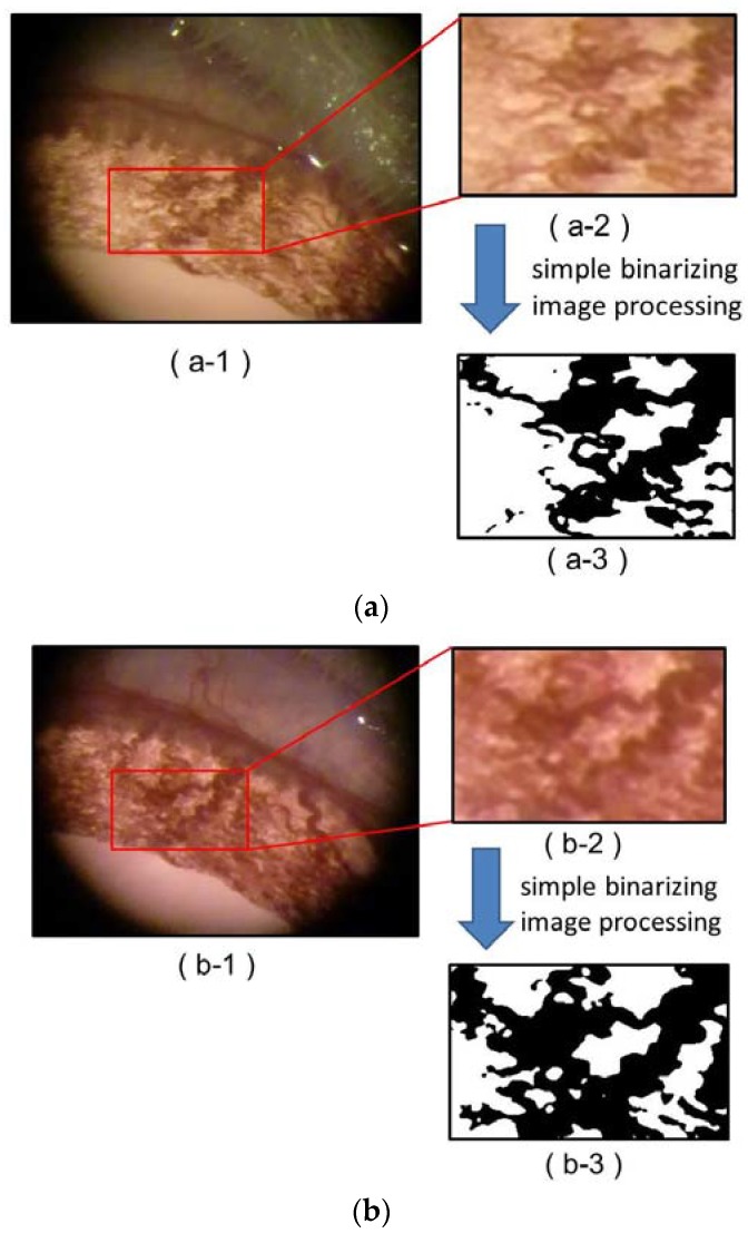
Figure 4.
Microvessels of a rat’s iris viewed under a magnifying glass before and after an injection of Hb-Vs [1] (a-1) Before the injection; (a-2) The specific area in the rat’s iris before the injection; (a-3) Two-dimensional changes in the microvessels were analyzed by area extraction using simple binarizing image processing (Image J, National Institutes of Health, Maryland, USA); (b-1) After the injection of Hb-Vs, showing a marked expansion in the microvessels, with the microvessel area in the bird’s-eye view increasing by 20%; (b-2) The specific area after the injection; (b-3) Two-dimensional changes in the microvessels were analyzed by area extraction using simple binarizing image processing.

