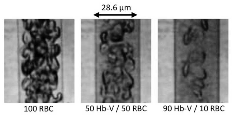
Figure 5.
Flow patterns of red blood cells (RBCs) mixed with Hb-Vs suspended in human serum albumin in a narrow tube (28.6 µm in diameter). The Hb-V solutions were mixed with the RBC suspension at volume ratios (Hb-V/RBC) of 0:100, 50:50 and 90:10. The Hb-V particles were homogeneously dispersed in the suspension medium but tended to become distributed in the marginal zone of the flow. The thickness of the RBC-free layer increased with increasing amounts of Hb-V, with the RBC-free phase becoming darker and semitransparent, indicating the presence of Hb-Vs. Hb concentration, 10 g/dl; centerline flow velocity, 1 mm/s [3,16].

