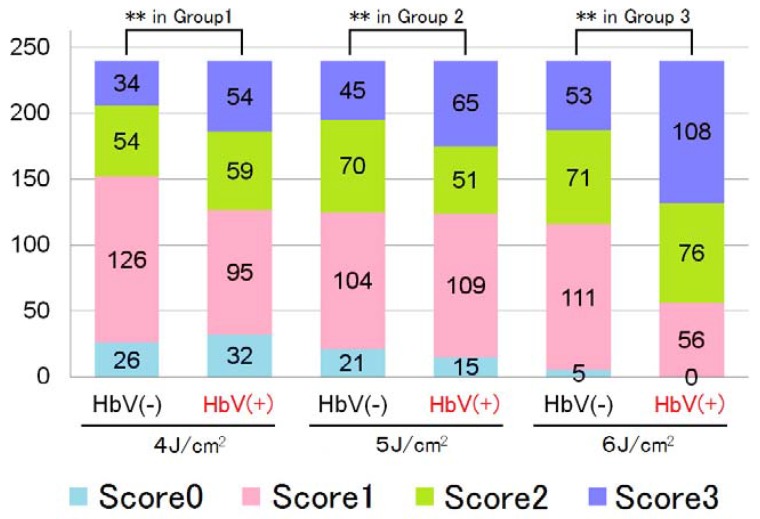
Figure 7.
Distribution of the histopathological findings scores after dye laser irradiation at three difference fluences in the chicken wattle subgroups with and without the administration of hemoglobin vesicles (Hb-Vs). ** P < 0.01 for the difference from the Hb-V(-) group (chi-square test). We irradiated a laser beam by a V-beam dye laser (Candera Corp., California, USA; wavelength, 595 nm; pulse width, 0.45–40 ms). No cooling device was used to protect the skin surface.

