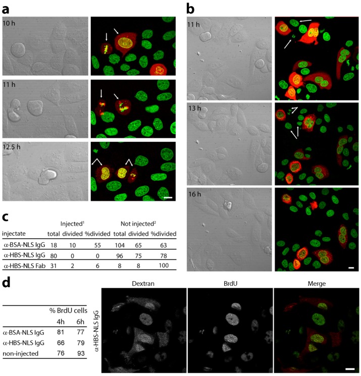
Figure 4.
Microinjected lamin A/C HBS antibodies blocked mitotic entry and delayed DNA replication. (a–c) Synchronized cells were injected within 1 h from release at G1/S with HBS or control antibodies conjugated to an SV40 NLS peptide using fluorescent dextran to identify injected cells. (a) Control antibody injected cells (αBSA) visualized from 6 h post-G1/S release (5 h post-injection). Images at times shown follow cells through mitosis with arrows. The control-injected cells went through mitosis at similar rates as non-injected cells in the same fields (c). (b) Lamin A/C HBS antibody-injected cells were followed longer as no cells entered mitosis in the timeframe of controls. (c) Numbers of cells followed for each condition, including also cells injected with Fab fragments of the lamin A/C HBS antibodies. Scale bars, 10 µm. (d) HBS antibody effects on DNA replication. HeLa nuclei were microinjected within 30 min after G1/S release and pulsed with BrdU starting at 3 h post-release for 1 h. The percentage of BrdU-positive cells at 4 and 6 h post-G1/S release for each condition is given in the table. To ensure that microinjection itself did not affect DNA replication, control antibodies (BSA-NLS) were injected into a parallel culture. A representative image of an injected, BrdU-labelled cell is shown.

