Figure 9.
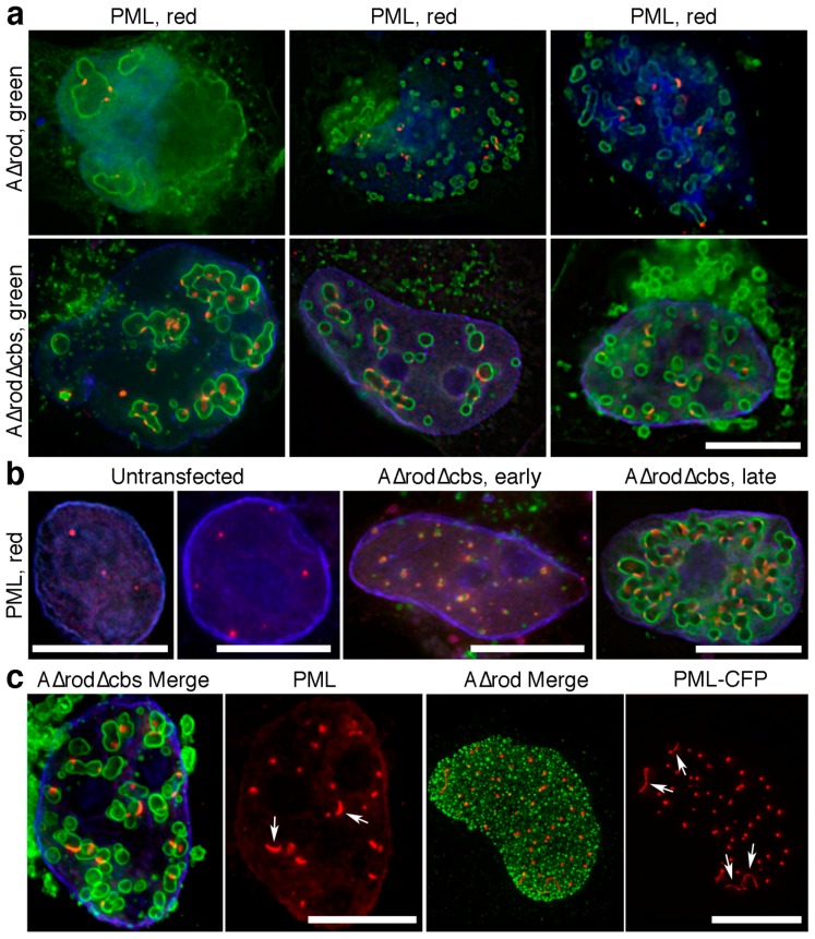
Potential PML interaction with lamin A. (a) HeLa cells expressing A∆rod and A∆rod∆hbs were stained with an antibody for PML. Nearly all PML foci were associated with the mini-lamin structures. Scale bar, 10 µm. (b) Numbers of observed PML foci increase in cells expressing A∆rod∆hbs. Left panels, untransfected cells exhibited relatively few PML foci. Middle panel, co-localization between A∆rod∆hbs and PML was observed even at early time points before the larger circular structures had formed. Right panel, later cells with fully-formed intranuclear A∆rod∆hbs circular structures exhibited both co-localization with PML and a considerable increase in the number of PML foci. Scale bars, 10 µm. (c) PML foci were often distended on the mini-lamin A structures. Left panels show an A∆rod∆hbs expressing cell with two more distended PML foci indicated by arrows that curve around the A∆rod∆hbs circular structures instead of appearing as a normal spot. Right panels show that, in HeLa cells co-expressing A∆rod and PML fused to CFP, extremely long distended PML structures (arrows) were observed, even in cells without significant development of the A∆rod circular structures. Scale bars, 10 µm.

