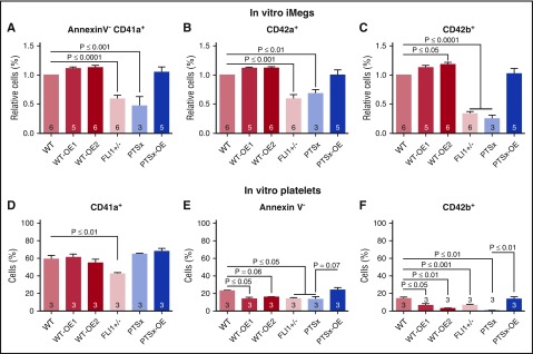
Figure 3.
Percentage of annexinV-CD41+CD42a+CD42b+ iMegs and in vitro–released platelets. Day 5 iMegs and released platelet-like particles were analyzed for surface markers, using flow cytometry. (A-C) iMegs were negative for annexin V and positive for CD41a, CD42a, and CD42b. (D-F) In vitro platelet-like particles positive for CD41a, negative for annexin V, and positive for CD42b. Means ± 1 SEM are shown with n = 3-6 independent experiments per arm. Significant P values were determined using 1-way ANOVA.

