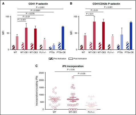
Figure 6.
Ex vivo and in vivo functional analyses of iPlts. (A) CD41+ and (B) CD41+CD42b+ iPlt mean fluorescence intensity (MFI) of surface P-selectin before and after thrombin stimulation of iPlts generated at 4 hours after iMeg infusion. Means ± 1 SEM are shown with n = 6 independent experiments per arm. Significance was determined by 1-way ANOVA. (C) Cremaster injuries were induced at 4 hours after iMeg infusion in NSG mice, and fluorescent images were recorded. The numbers reported are of human calcein AM–stained particles incorporated into a growing thrombus after normalization by dividing by the percentage of circulating CD42b+-human platelets as part of the total circulating platelets. Shown are the individual data point and mean ± 1 SEM of experiments from 4 individual mice with up to 6 injuries per mouse. Significance was determined by 1-way ANOVA.

