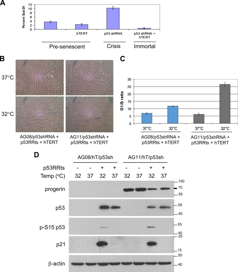
FIG 2.
Progerin-induced premature senescence is dependent on p53. (A) Determination of crisis (apoptosis) in AG11 HGPS cells expressing pBABE vector control (MPD 25), hTERT (MPD 45), p53 shRNA (MPD 49), or both hTERT and p53shRNA (MPD 45). Apoptosis was determined by flow cytometry on the basis of sub-G1 content after propidium iodide staining. At the time of analysis, MPD 49 p53 shRNA cells were in a state of decline in which overall cell numbers were decreasing, while all other cell lines were actively growing. (B to D) p53RRts and hTERT were coexpressed in normal AG08/p53 shRNA and HGPS AG11/p53 shRNA. The cells were assessed at an MPD of 60 (AG08/p53sh/p53RRts/hTERT) or MPD of 50 (AG11/p53sh/p53RRts/hTERT). hTERT was expressed so that the contribution of progerin could be assessed independently of telomere erosion during premature senescence. (B) Cell growth was assessed at 37°C and at 32°C. Cells were visualized after β-galactosidase staining. hTERT-p5RRts-expressing AG11 cells at 32°C were 82% ± 5.6% positive β-galactosidase, demonstrating telomere-independent senescence. (C) The cells used for panel B were stained with propidium iodide, and the G1/S ratios were obtained from the cell cycle profiles using flow cytometry. Values shown represent the means and SEM (n = 3). (D) Western blot analysis confirming p53 knockdown and p53RRts and progerin expression in the cells used for panel B. The activation of p53 at 32°C is demonstrated by the upregulation of p21 and by the phosphorylation of p53 at Ser15.

