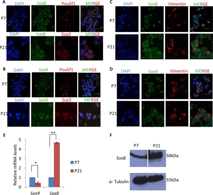
FIG 1.
Expression of Sox8 and Sox9 in mouse testes. The Sox8 and α-tubulin Western blots are from different gels. For the Sox8 blot, the space separating P7 and P21 is due to a protein marker lane that was excised. (A and B) Expression of Sox8 (A) and Sox9 (B) in 7-day-old mouse testis with Pou5f1 as a marker for spermatogonial cells and in 21-day-old mouse testis with Scp3 as a marker for spermatocytes. (C and D) Expression of Sox8 (C) and Sox9 (D) in 7-day-old and 21-day-old mouse testis with vimentin as a marker for Sertoli cells. (E) Expression analysis of Sox8 and Sox9 in P7 and P21 mouse testes. (F) Western blot showing Sox8 expression in P7 and P21 mouse testes. Data in panel E are plotted as means ± standard deviations (SD) (n = 4). **, P ≤ 0.01; *, P ≤ 0.05 (two-tailed Student's t test). Bars, 25 μm.

