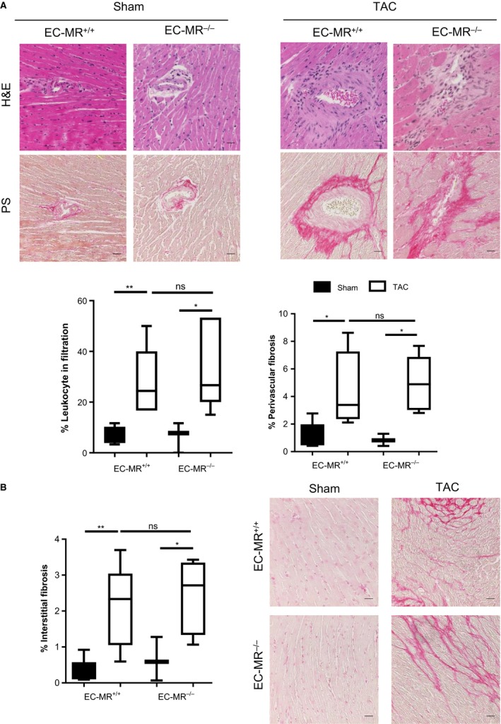
Figure 4.

EC‐MR does not regulate perivascular inflammation, perivascular, and interstitial collagen deposition in response to TAC. (A) Representative H&E and Picrosirius red‐stained LV sections, showing areas with elevated leukocyte infiltration colocalizing with areas presenting high percentage of perivascular fibrosis; quantifications of infiltrates and perivascular collagen deposition are shown below. (B) LV interstitial collagen deposition quantified as percent fibrosis on the left. Scale bar 25 μm. n = 5 EC‐MR +/+ Sham, 8 EC‐MR +/+ TAC, 3 EC‐MR −/− Sham, 8 EC‐MR −/− TAC. Statistics D'Agostino & Pearson normality test, followed by unpaired Student's t‐test
