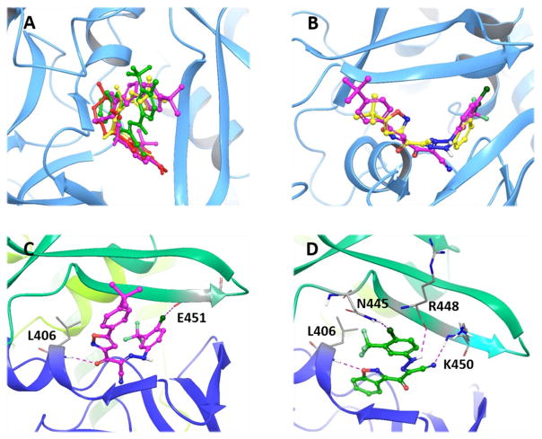
Fig. 4.
(A) Overlay analysis of molecular docking poses of 1, 10 and 33 binding at the cAMP binding domain B (CBD-B) of EPAC2 protein (PDB Code 3CF6). cAMP is shown in red, 1 in yellow, 10 in magenta, and 33 in green. (B) Overlay of molecular docking poses of 1 (yellow) and 10 (magenta) binding at the CBD-B of EPAC2. (C) Predicted binding mode of 10 docked into the CBD-B of EPAC2. 10 is shown in magenta ball and stick representation. Key residues are displayed in sticks. Hydrogen bonds and halogen bond are shown in dotted purple lines. (D) Predicted binding mode of 33 docked into the CBD-B of EPAC2. 33 is shown in green ball and stick representation. Key residues are displayed in sticks. Hydrogen bonds and halogen bond are shown in dotted purple lines.

