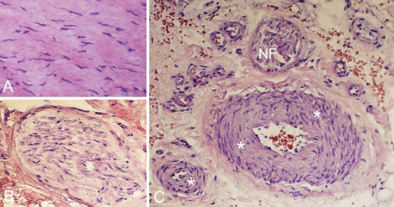
Figure 2.

Sections stained for morphology (H&E). The tendinous tissue exhibits large numbers of tenocytes with different shapes including rounded and wavy appearance (A). There are nerve structures such as nerve fascicles (B) and also large numbers of blood vessels C). Asterisks point on blood vessel walls; NF indicates nerve fascicles.
