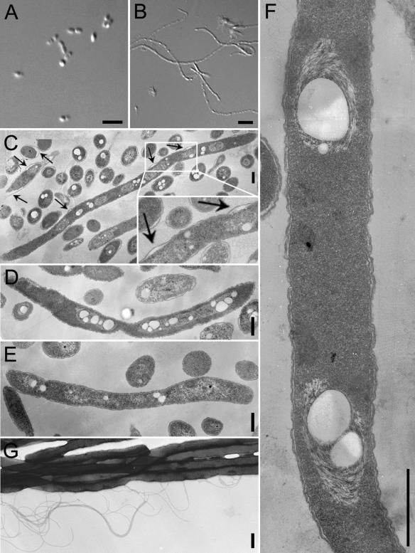
FIG. 1.
Phase-contrast and TEM of antibiotic-induced B. pseudomallei filaments. (A and B) Phase-contrast microscopy pictures of B. pseudomallei not treated with antibiotics (A) or treated with 0.5 μg ceftazidime per ml (B) for 16 h; (C to E) TEM pictures of thin sections of B. pseudomallei filaments induced by 0.5 μg of ceftazidime per ml (C), 4 μg of ofloxacin per ml (D), or 8 μg of trimethoprim per ml (E) for 16 h; (F) a higher-magnification picture of filaments induced by 0.5 μg of ceftazidime per ml; note the absence of any visible septa in the filament; (G) TEM picture of filaments induced by 0.5 μg of ceftazidime per ml without sectioning, which shows the presence of flagella. The arrows in panel C indicate points of periplasmic space enlargement. Bars in panels A and B, 5 μm; bars in panels C to G, 500 nm.

