
Figure 4.
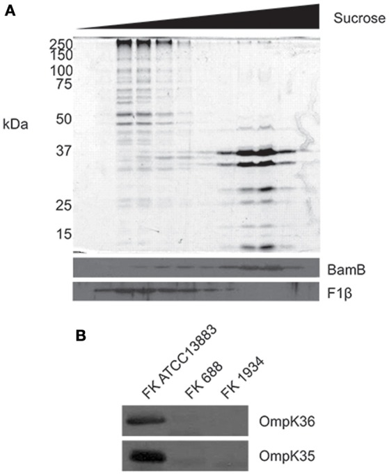
Remodeling of the outer membrane proteome in the XDR K. pneumoniae isolates. (A) Total membranes were isolated from K. pneumoniae ATCC 13883 and subjected to sucrose density fractionation. Membrane fractions were subject to SDS-PAGE, and then Coomassie blue staining (top panel) and immunoblotting (bottom panel) using antisera recognizing known outer (BamB) and inner (F1β) membrane proteins. (B) Whole cell lysates from K. pneumoniae ATCC 13883 and K. pneumoniae clinical isolates FK688 and FK1934 were analyzed by SDS-PAGE and western immunoblotting for OmpK35 and OmpK36.