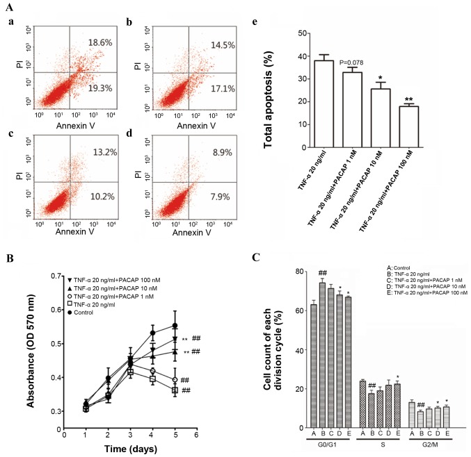
Figure 3.
PACAP restores TNF-α-induced apoptosis in ECFCs. (A) Effect of TNF-α and PACAP on apoptosis of ECFCs. The cell populations in early and late apoptosis were determined using flow cytometry: (Aa) 20 ng/ml TNF-α; (Ab) 20 ng/ml TNF-α + 1 nM PACAP; (Ac) 20 ng/ml TNF-α + 10 nM PACAP; (Ad) 20 ng/ml TNF-α + 100 nM PACAP; (Ae) Bar chart of ECFC apoptosis. (B) Cell proliferation was evaluated by MTT assay. PACAP partially restored proliferation of TNF-α-treated ECFCs. (C) Proportion of cells in each cell cycle phase determined by flow cytometry. ##P<0.01 vs. Control group; *P<0.05 and **P<0.01 vs. 20 ng/ml TNF-α group. PACAP, pituitary adenylate cyclase-activating polypeptide; TNF-α, tumor necrosis factor-α; ECFC, endothelial colony-forming cell; PI, propidium iodide.

