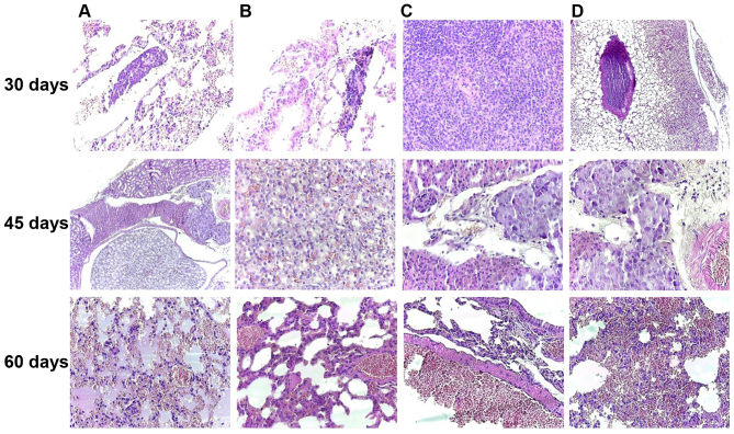
Figure 8.
Melanoma achromic metastasis at 30 days post-inoculation in the lung (A and B, original magnification, ×40) and spleen (C, original magnification, ×100) and tumor thrombus in the big arterial blood vessel (D, original magnification, ×40); Kidney metastases was noted in the 45th day after inoculation, disposed between the renal tubules and nearby medium-sized arterial vessels of the renal medulla (A, original magnification, ×40) and composed of achromic epithelioid cells similar to those observed in the primary tumor (B, original magnification, ×100; C and D, original magnification, ×40); On the 60th day of the experiment, the most affected internal organ was the lung, with hemorrhagic alveolitis (A, original magnification, ×40), hyperemia of small- and medium-sized blood vessels (B and C, original magnification, ×100) and many extravasated erythrocytes in the alveolar spaces (D, original magnification, ×40), H&E staining.

