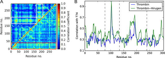
Fig. 2.

(A) Comparison of generalized correlations in isolated thrombin (lower triangle) and thrombin–hirugen (upper triangle). The prominent role of the light chain in isolated thrombin is outlined by the red (dark gray in the print version) box, and the global increase of correlation with the 148CT Loop in thrombin–hirugen is highlighted by white boxes. (B) The pairwise correlations between thrombin residues and Exosite I residue T74. The binding of hirugen raises correlations with T74 system wide, but the catalytic residues (vertical dotted lines) experience a significant increase in correlation strength.
