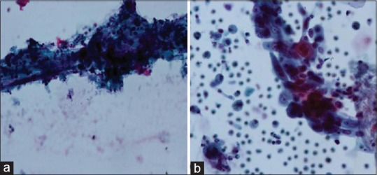
Figure 1.

Comparison of CC and LBC in BAL specimen in cases of squamous cell carcinoma. (a) CC preparation shows atypical keratinised cellls entrapped in inflammatory exudate. (Pap stain x600), (b) LBC preparation shows atypical squamous cells with preserved nuclear and cytoplasmic details in a cleaner background (Pap stain x600)
