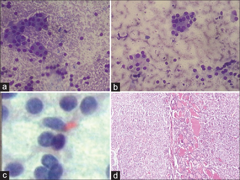
Figure 1.

(a and b) FNAC thyroid shows mostly Hurthle cell change with few small follicles and abundant colloid in background (a: May–Grunwald–Giemsa; ×100, b: May-Grünwald-Giemsa; ×200); (c) Smears show few nuclei showing intra nuclear inclusions. Case was diagnosed as category AUS/FLUS of BSRTC (Pap stain x1000); (d) On histopathological examination, the case was diagnosed as follicular variant of papillary carcinoma (H and E stain x100)
