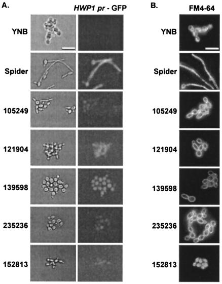
FIG. 3.
Effects of small molecules on hyphal gene expression and endocytosis. (A) KTCa1 cells in Spider medium were added to 384-well microplates with an optical-glass bottom. Induction of HWP1 expression was determined after 4 h of incubation. (B) To stain the vacuoles of SC5314 cells, the assay was carried out in optical-glass-bottomed microplates for 3 h, then FM4-64 was added, and the plate was returned to 37°C for 30 min. The medium containing FM4-64 was then aspirated out of each well, and 100 μl of fresh medium was added. The plate was returned to 37°C, and digital images of the cells were captured at 120 min with a 100× objective for wells containing budded cells or a 60× objective for wells containing hyphal cells.

