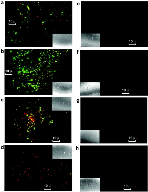
FIG. 4.
Cells treated with PMC-EF (a to d) or medium alone (e to h) stained with propidium iodide and FITC-labeled antibody and examined under a confocal microscope. The following times elapsed after additions: 0.1 h, panels a and e; 0.5 h, panels b and f; 1 h, panels c and g; and 2 h, panels d and h. Insets show full-field views.

