Fig. 2.
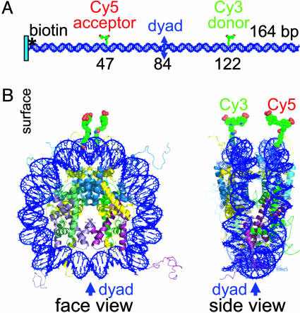
Locations of the donor and acceptor fluorophores on nucleosomal 164-bp DNA. (A) Naked DNA. Marked DNA bases are sites for attachment of acceptor and donor dyes, bases 47 and 122, respectively, on separate strands of DNA. The nucleosomal pseudodyad is at position 84. The distance between the methyl carbons of the two labeled pyrimidines located on opposite strands of the linear DNA is 75 bp (25 nm), but only ≈3 nm (from DNA gyre to DNA gyre) in the particle. This placement of the dyes should result in no single-pair FRET on the linear DNA fragment and high-efficiency energy transfer in the nucleosome. (B) Face and side views of the nucleosome. Modeling of Protein Data Bank structure 1KX5 (4) was performed by using xfit (41), and the model was rendered with pymol (42). We thank J. Harp for help with this figure.

