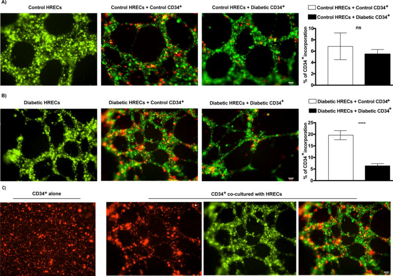
FIG. 3. Reduced incorporation of diabetic CD34+ CACs into diabetic HRECs tubes.

Tube formation by HRECs (Qtracker 525, green) isolated from control (A) or diabetic (B) donors either without CACs (left panel), or co-incubated with control (middle panel) or diabetic (right panel) CACs (Qtracker 655, red) is shown. Quantification of % of CD34+ CACs incorporation into HRECs tubes is shown on far right. Data are means ± SEM (n= 4−7). *** P < 0.0001, significantly different from control as determined by Student’s t -test; not significant at P > 0.05. Scale bar = 50 μm. (C) CD34+ CACs alone were not able to form tube-like structures (left panel), but incorporated into HREC tubes, forming tube-like structures, when co-cultured with HRECs (right three panels). Abbreviations: CACs, circulating angiogenic cells.
