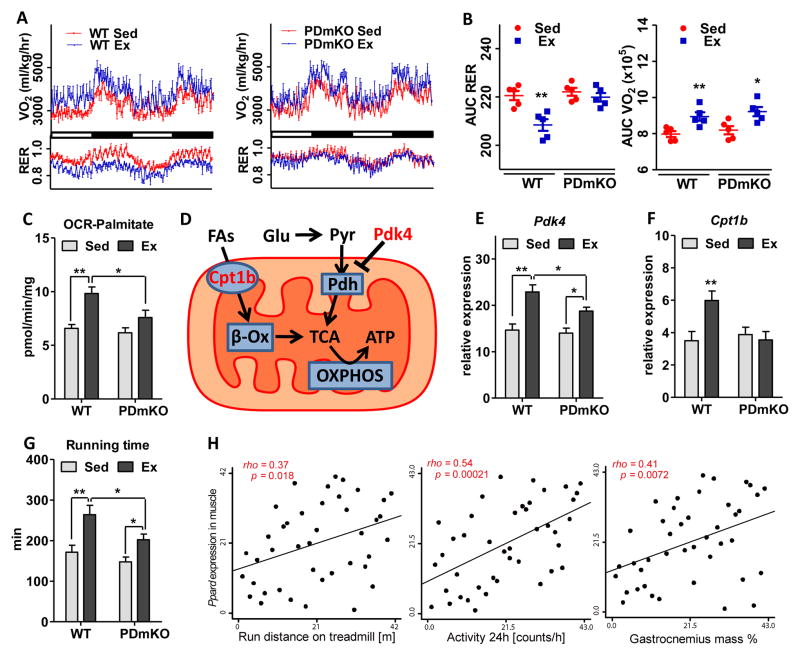
Figure 1. Exercise induces a PPARδ-dependent shift in muscle energy substrate utilization.
Experiments were performed in the same set of 4-month-old WT or PDmKO mice with or without 4 weeks of exercise training (n=5). (A) Oxygen consumption rate (OCR, VO2) and respiratory exchange ratio (RER, VCO2/VO2) measured over a 48-hour period. (B) Quantitative analysis of VO2 and RER using area under curve (AUC) of the data in (A). (C) OCR using palmitoyl-carnitine as the substrate in freshly isolated mitochondria from quadriceps muscle. (D) Diagram showing the two major types of fuel sources (glucose and FAs) and the gatekeeper enzymes that control their mitochondrial influx. (E–F) mRNA expression levels of Cpt1b and Pdk4 in soleus. (G) Total running time in a run-to-exhaustion endurance test. (H) Correlations between PPARδ expression in quadriceps and running distance, activity, and muscle mass. Asterisks denote statistically significant differences (*p < 0.05, **p < 0.01).

