
Figure 2. MiR-155−/− Mice Infected with LRV1+ L.g Parasites Had Decreased Disease Pathology when Compared to WT Mice.
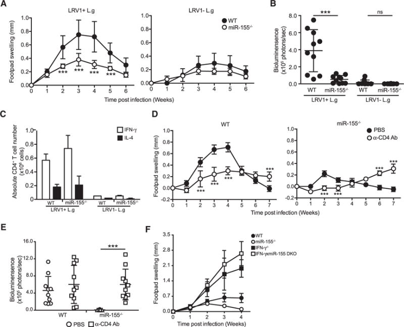
(A and B) Hind footpads of WT and MiR-155−/− mice were infected with 1 × 10⁶ LRV1+ L.g or LRV1 − L.g.
(A) The graph displays the weekly measurement of footpad swelling.
(B) Parasite burden was determined at 3 weeks after infection by bioluminescence imaging.
(C) Popliteal lymph nodes were collected from WT and miR-155 mice infected with LRV1+ L.g or LRV1 − L.g on the footpad after 3 weeks of infection. Intracellular FACS analysis was performed using primary ex vivo lymphocytes extracted and stimulated with PMA/ionomycin to measure IL-4 and IFN-γ levels in CD4+ T cells (see also Figure S2).
(D and E) Mice were intraperitoneally injected with CD4-depleting antibody 5 days prior to LRV1+ L.g infection and then on a weekly basis for 6 weeks. A group of mice was treated with PBS as a negative control (see also Figures S3 and S4).
(D) Footpad swelling of LRV1+ L.g-infected WT and MiR-155−/− mice.
(E) Parasite load was measured by bioluminescence 4 weeks post-infection.
(F) The measurement of footpad swelling in WT, MiR-155−/−, IFN-γ, and IFN-γ xmiR-155 DKO mice following of LRV1+ L.g infection.
Data show mean ± SD from representative experiments (n = 4–5 mice) of two (C–E) or three (A, B, and F) independent experiments. Each dot in the scatter blot represents a footpad (B and E). Statistical significance is calculated using two-way ANOVA analysis with Bonferonni’s post test (A and D) and unpaired Student’s t test (B and E). Not significant (NS), *p < 0.05, and ***p < 0.001. See also Figure S1.