
Figure 3. MiR-155−/− Macrophages Exhibited Similar Proinflammatory Cytokine Response Compared to WT Macrophages in Response to LRV1+ L.g or Poly I:C.
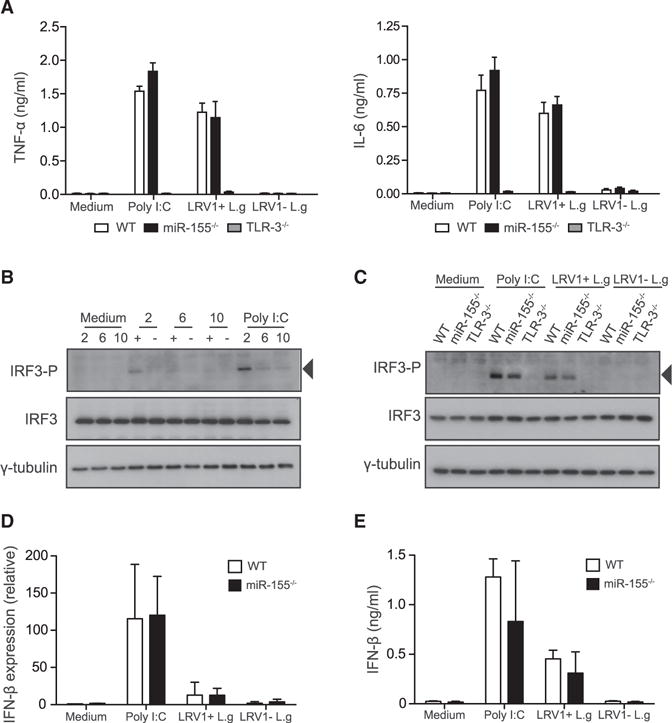
(A–E) BMMs were infected with LRV1+ L.g (+) or LRV1 − L.g (). Cells were treated with medium or poly I:C (2 μg/ml) as a control for indicated times.
(A) The cell-free culture supernatant was collected 24 hr post-treatment from treated macrophages derived from WT, TLR-3−/−, and miR-155−/− mice. These samples were assayed for IL-6 and TNF-α using ELISA.
(B) Total cell lysate of treated WT macrophages was analyzed by western blot with IRF3-P, IRF-3, and γ-tubulin after 2, 6, and 10 hr infection.
(C) WT, TLR-3−/−, and miR-155−/− BMMs were lysed after 2 hr and immunoblotted for the indicated antibodies.
(D) IFN-β mRNA levels were assayed after 2 hr incubation using RT-PCR. Values were normalized using L32. Transcript levels were calculated relative to unstimulated WT macrophages.
(E) Protein level of IFN-β was quantified in collected cell culture medium after 6 hr treatment. Data are mean ± SD from three pooled independent experiments. Representative blots were shown from three independent experiments.