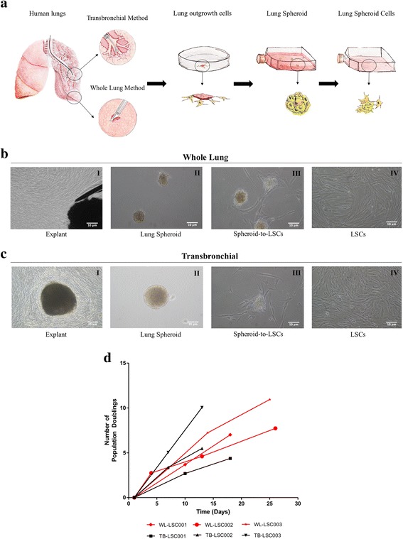
Fig. 1.

Growth potential lung spheroid cells derived from whole lung and biopsy specimens. a: Schematic showing the protocol to derive lung spheroid cells (LSCs). b I-IV: Bright field image of each stage of WL-LSC generation. b(I): Explant tissue in the middle surrounded by outgrowth of cells. b(II): Lung spheroids formed from explant-derived cells (EDCs). b(III): Plated spheres onto fibronectin coated surface allowing lung spheroid cells to grow out from the spheroids. b(IV): Expansion of LSCs in adherent culture. c I-IV: Bright field image of each stage of TB-LSC generation. d: Cumulative population doubling for TB-LSCs and WL-LSCs from three different donors. Scale Bar = 10 μm
