Fig. 3.

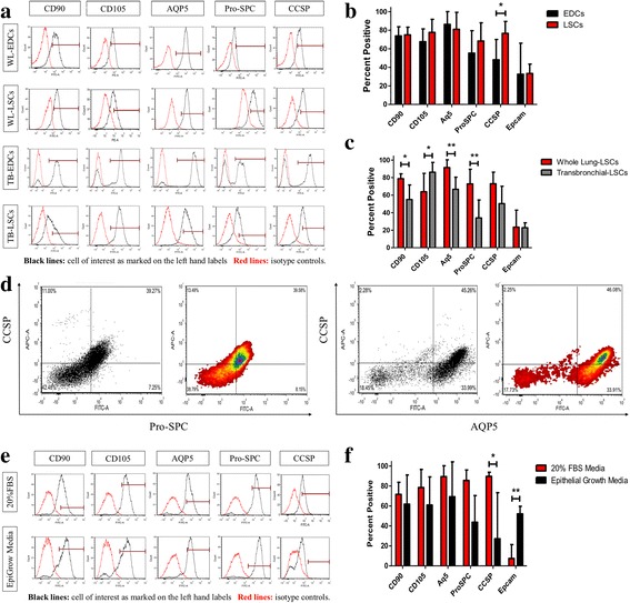
Phenotype Analysis of lung spheroid cells. a: Representative flow cytometry histograms of WL-EDCs, WL-LSCs, and TB-LSCs for expression of CD90, CD105, AQP5, Pro-SPC and CCSP. Black lines in the histograms are the cell of interest as marked on the left hand labels. Red lines in the histograms are the unstained or isotype controls. b-c: Cumulative data for the expression CD90, CD105, AQP5, Pro-SPC, CCSP and Epcam showing the expression change between b) WL-EDCs to WL-LSCs (n = 5–12) and c) WL-LSCs and TB-LSCs (n = 7-12). d: Double stained flow cytometry plot of CCSP verse ProSPC and CCSP verse AQP5. e: Representative flow cytometry histograms of WL-LSCs in either IMDM with 20% FBS or epithelial cell growth medium for expression of CD90, CD105, A15, Pro-SPC, and CCSP. Black lines in the histograms are the cell of interest as marked on the left hand labels. Red lines in the histograms are the unstained or isotype controls. f: Cumulative data for the expression CD90, CD105, A15, Pro-SPC, CCSP, and Epcam (n = 3-4). Data represented as mean ± SD. * p < 0.05; **p < 0.01
