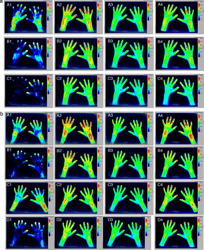
Fig. 2.

a FOI of a patient treated with adalimumab. Row A, baseline; row B, week 12; row C, week 24; column 1, phase 1; column 2, phase 2; column 3, phase 3; column 4, composite image. At baseline, signals detected were grade 3 over PIP 3 of the right hand in phases 1 and 2 and composite image. Left-hand PIP 2–5 showed grade 2 signals in phase 2 and grade 1 signal in phase 3 and composite image. Left-hand PIP 1 showed grade 2 in phase 2, grade 0 in phase 3 and grade 1 in composite image. After 12 weeks of treatment, improvement of signal intensity was visible. Especially, right-hand PIP 3 still shows grade 3 signal in phase 2. Further reduction of signaling was noted at week 24. b FOI of a patient with polyarticular JIA who started etanercept treatment. Row A, baseline; row B, week 12; row C, week 24; row D, week 52; column 1, phase 1; column 2, phase 2; column 3, phase 3; column 4, composite image. At baseline, both wrists, both side PIP 2–5 and DIP 2–4 showed grade 3 signals in phase 2 but grade 1 in phase 3. Both DIP 5 showed weaker signals. Twenty-four-week imaging, especially in phase 2, demonstrates subclinical activity. At clinical examination the patient already had no active joints. Upon constant clinical remission on treatment, there is further improvement of FOI findings visible after 1 year
