
Figure 4.
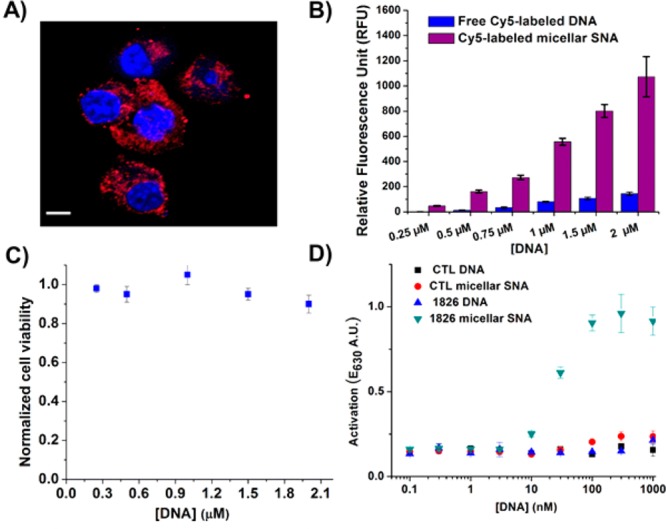
(A) A confocal fluorescent micrograph of HEK-Blue mTLR9 cells that were incubated with Cy5-labeled micellar SNAs ([DNA] = 100 nM) for 4 h. Cell nuclei were stained with Hoechst 33342 (scale bar = 20 μm). (B) Flow cytometry analysis of HEK-Blue mTLR9 cells that have been incubated with free Cy5-labeled DNA (blue bars) and Cy5-labeled micellar SNAs after 16 h (purple bars), showing a higher fluorescence intensity for the latter. (C) A cell-viability assay for HEK-Blue mTLR9 cells after treatment with micellar SNAs for 24 h. (D) Plots of the amounts of secreted alkaline phosphatase (SEAP) by HEK-Blue cells, as visualized by a colorimetric assay, showing enhanced immunostimulatory activity by micellar SNAs in comparison to control micellar SNAs bearing a T20 sequence and unmodified linear nucleic acids.