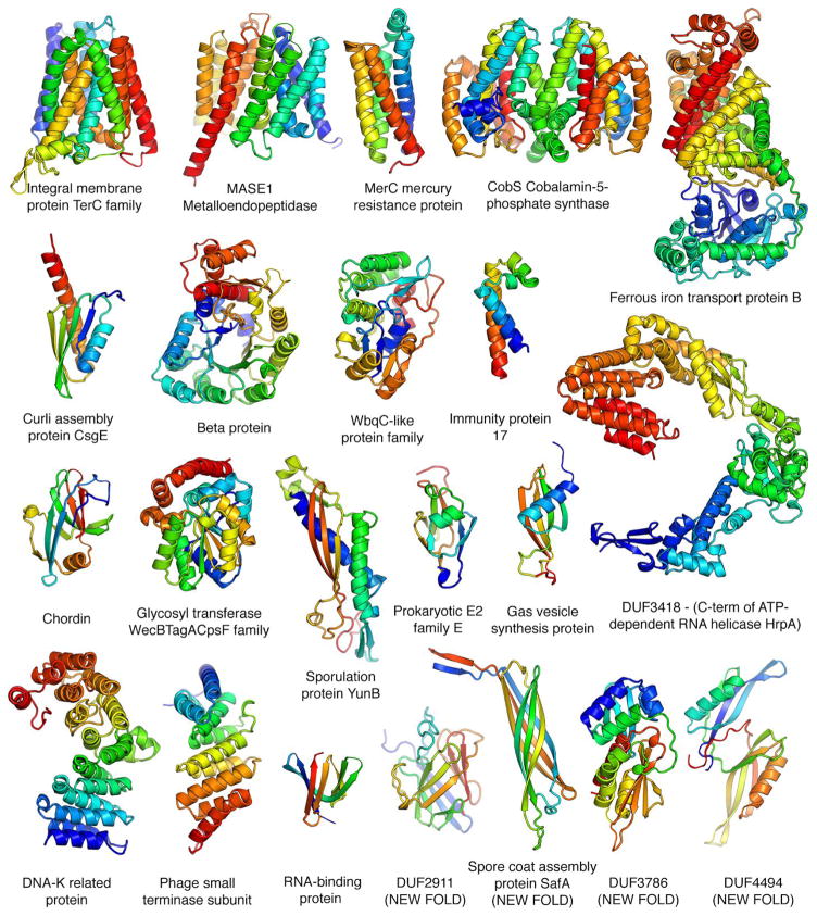
Fig. 3.
Representative structure models for selected PFAM families. Membrane proteins are on the top row; new folds on the bottom right. The multidomain models of the iron transporter and RNA helicase and the dimeric model of CobS, an enzyme in vitamin B synthesis, are guided by both intra- and inter-chain coevolution restraints.

