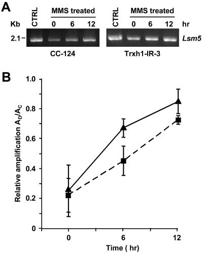
FIG. 5.
Repair of MMS-induced DNA lesions in the wild type (CC-124) and a Trxh1 RNAi epi-mutant (Trxh1-IR-3) as examined by semiquantitative PCR. (A) Amplification of a 2.1-kb genomic DNA fragment (Lsm5) by using, as the template, DNA isolated from cells immediately after treatment with 10 mM MMS (0 h) or after allowing the cells to recover for 6 or 12 h in the absence of MMS. Untreated control cells (CTRL) were also analyzed. The amplified products were resolved by agarose gel electrophoresis and stained with ethidium bromide. (B) Relative amplification of the 2.1-kb fragment calculated by dividing the amount of amplification from damaged samples (AD) by the amount of amplification from nondamaged controls (AC). Each graph point represents the mean (± standard error) of results of three independent experiments. Symbols: ▴, CC-124; ▪, Trxh1-IR-3.

