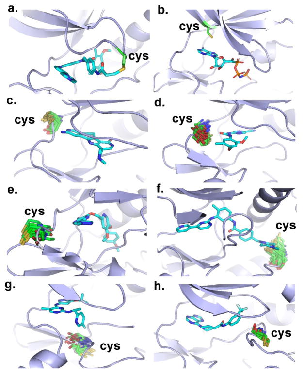
Figure 4.
Cysteines at different locations; sulfur atom in yellow, cysteine in green; and ligand in blue. (a) Cysteine at P-loop (pdb id 4r6v). (b) Cysteine at roof of the binding pocket (pdb id 4riy). (c) Cysteines at location Hinge-1 of the hinge region (representative pdb id 3m2w). (d) Cysteines at location Hinge-2 of the hinge region (representative pdb id 2ywp). (e) Cysteines at location Hinge-3 of the hinge region (representative pdb id 3lco). (f) Cysteines at location Catalytic-2 of the catalytic loop (representative pdb id 1t46). (g) Cysteines at location Catalytic-9 of the catalytic loop (representative pdb id 4a0j). (h) Cysteine at location DFG-4 close to the DFG peptide (representative pdb id 3wf7).

